

An exciting technique in mammography, Contrast-Enhanced Mammography (CEM) is an increasingly important and effective method providers are using to detect breast health issues in patients with dense breast tissue and other extenuating factors. At MagView, we’re dedicated to helping breast health centers improve service and outcomes through cutting-edge technology that allows for higher accuracy and simpler, more effective standardized reporting.
What is Contrast-Enhanced Mammography (CEM)?
Contrast-Enhanced Mammography (CEM) is a technique in breast health imaging that uses intravenous contrast agents in tandem with traditional mammography, which allows providers to get a more detailed, accurate view of any breast abnormalities a patient might have. This technique is especially useful in patients with dense breast tissue.
Each mammography technique holds an important place in breast health care, with CEM holding special importance in the realm of dense breast tissue, and determining the severity of breast cancer cases. It is also typically quicker and more cost-effective than an MRI.
How Contrast-Enhanced Mammography Works
CEM begins with intravenous (IV) injection of an iodine-based contrast agent, which travels through the blood stream for about two minutes before imaging starts. This allows providers to get a clearer picture of breast symptoms, abnormalities, and the progression of any cancer, if detected. Patients often report a brief feeling of full-body warmth, the feeling of needing to urinate, and a metallic taste in their mouth.
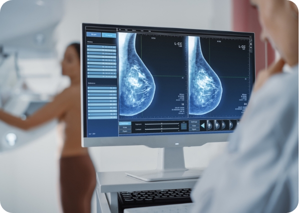
As mentioned above, CEM is the use of both intravenous contrast agent and traditional mammography. After the contrast agent has been given enough time to spread throughout the body, imaging, or a traditional digital mammogram, will begin.
Two images of each breast are taken in two positions, resulting in four images per breast. The first image is taken like a standard mammogram, with “low energy” x-rays, and the second images are taken using higher energy x-rays. When the images are compared, providers are able to see abnormalities that weren’t visible without the contrast agent.
The growing popularity of CE Mammography
Contrast-enhanced mammography has quickly grown in popularity over the past few decades; so much so that it is being compared in effectiveness to MRI when it comes to breast cancer diagnostics. According to recent research, “Contrast-enhanced mammography (CEM) is becoming a widely adopted modality in breast imaging over the past few decades and exponentially so over the last few years, with strong evidence of high diagnostic performance in cancer detection. Evidence is also growing indicating comparative performance of CEM to MRI in sensitivity with fewer false positive rates.”
These strides in technology are, ultimately, an incredible benefit to the study of breast cancer and breast health, but as CEM increases in popularity, so, too, does the need for standardization of reporting best practices. In fact, CEM is becoming so popular that, in 2022, the American College of Radiology (ACR) published a supplement to ACR BI-RADS® Mammography 2013 that specifically addresses the need for universality in reporting:
“Contrast enhanced mammography (CEM) was first approved by the Food and Drug Administration (FDA) in 2011 and its use is increasing,” ACR says. “CEM is more sensitive than mammography or ultrasound for the detection of malignancy. Given that utilization is increasing, it is important that a lexicon be available to allow for consistency in reporting and also to allow for validation of standardized terms through studies looking at the performance of CEM in a variety of clinical settings. The next edition of BI-RADS® is under development but rather than waiting to issue a section on CEM until the release of the new edition, this supplement is available to facilitate the interpretation and reporting of CEM studies.”
Key Benefits of CEM Reporting
The benefits of CE Mammography are becoming more and more clear, which is naturally increasing the use of this technique in breast health centers. This increases the need for accurate, efficient reporting of these results, so that interpretation and treatment can be consistently executed and documented.
Some key benefits of standardized CEM Reporting include:
- Encouraging consistency and uniformity in radiography documentation, specifically in complex areas like breast cancer
- Clarity in enhanced mammographic reporting, which removes ambiguous, inconsistent language and helps providers interpret results accurately
- Accuracy in documentation, making reports easier to access for auditing and review
- Improved patient safety and outcomes through efficiency and improved accuracy
Interpreting Findings & Diagnostic Considerations
The BI-RADS® (Breast Imaging Reporting and Data System) is a standardized reporting framework designed to help breast health centers embrace a unified method of reporting. The Contrast-Enhanced Mammography (CEM) Lexicon was published in April 2022 to act as a supplement to the BI-RADS® CEM, improving accuracy as the popularity of Contrast Enhanced Mammography increased.
These frameworks ultimately work toward improved clinician communication between referring physicians and radiologists, allowing for more consistent imaging interpretations and improved diagnostic accuracy.
Optimize Mammography Workflow with CEM Reporting with MagView
MagView is well-known as the leading provider of solutions for breast center information management. As a result, we understand the critical importance of effective data integration to optimize the mammography workflow — which is why CEM reporting is now a critical part of MagView’s breast health solutions. Our structured reporting ensures reports that are concise, consistent, and compliant with regulatory requirements.
Are you ready to improve consistency, efficiency, and accuracy in your center’s CEM reporting techniques? Contact MagView today to schedule your free demo.
Resources:
- https://borderradiology.com.au/the-importance-of-standardised-reporting-for-accurate-radiologic-diagnoses/
- https://www.gehealthcare.com/-/media/GEHC/US/Files/Products/Mammography/Contrast-Enhanced-Mammography-Lexicon?srsltid=AfmBOooNplgnDHtBSClHj0SmSy0kTz0n7NwfFF50iB2lO2eYXiaDbGwc
- https://cancerimagingjournal.biomedcentral.com/articles/10.1186/s40644-023-00526-1


 logo")


")


")


![shutterstock_1299510538-[Converted]](https://azuretest.magview.com/wp-content/uploads/2023/05/shutterstock_1299510538-Converted.jpg "monitoring breast density")